
Budhi Arifin Noor, Dion Ade Putra, Oktaviati, Ridho Ardhi Syaiful, Rizky Amaliah, Ira, Bob Andinata
Bedah Umum, Departemen Ilmu Bedah, FKUI/RSCM, Jakarta, Indonesia, Januari 2011
ILUSTRASI KASUS
Perempuan, 35 tahun dengan benjolan di leher kiri yang semakin membesar sejak 15 tahun.
Pada pemeriksaan fisik didapatkan tanda vital normal. Status lokalis di leher depan terdapat massa berukuran 4,5 x 4 x 1 cm, konsistensi padat, dapat digerakkan, berbatas tegas, berlobul dan ikut bergerak ketika menelan.
Pemeriksaan laboratorium didapatkan: T3: 1,7 nmol/L, fT4: 0,51 pmol/L, TSH: 9,21 uIU/mL. Pemeriksaan USG tiroid mendapatkan gambaran nodul multipel di tiroid kiri. Pada thyroid scan didapatkan kesan nodul multipel dengan aktivitas campuran dan thyroid uptake dalam batas normal. Pemeriksaan patologi anatomi melalui Fine Needle Aspiration Biopsy (FNAB) tidak ditemukan sel ganas.
Pada pasien ini ditegakkan diagnosis kerja Struma Nodosa Non Toksik (SNNT) sinistra suspek jinak dan direncanakan untuk menjalani operasi lobektomi per-MIVAT + Vriescope (VC).
Pasca-operatif pasien dirawat di ruang rawat biasa. Selama perawatan, kondisi stabil, produksi drain menurun. Hari ketiga, drain dilepas dan pasien diperbolehkan pulang. Pengobatan dilanjutkan di poliklinik bedah tumor RSCM.
 |
| Foto pre op anterior |
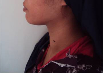 | ||||
| foto pre op lateral |
 |
| gambar scan tiroid 1 |
 |
| gambar scan tiroid 2 |
 |
| gambar usg 1 |
 |
| gambar usg 2 |
OPERATIF
Dilakukan insisi pada superolateral mammae sinistra (sekitar midaksilaris) untuk port pertama mengarah ke sternal notch kemudian kamera dimasukkan. Dilakukan insisi pada daerah aksila kiri tepi lateral m. pektoralis mayor, dimasukkan trokar no. 5 ke arah ujung trokar pertama. Dilakukan insisi port ketiga pada areola mammae sinistra arah jam 11, kemudian dimasukkan trokar no. 5 ke arah ujung trokar pertama. Kemudian dilakukan pembebasan dengan memisahkan subkutis dengan otot ke arah sternal notch, identifikasi m. sternokleidomastoideus, dibuka, dan identifkasi m. omohioid, dibuka, identifikasi m. sternotiroid, dibuka, identifikasi kelenjar tiroid. Dilakukan pembebasan kelanjar tiroid dengan harmoni skalpel, dimulai dari bagian superior kemudian media dan inferior dengan mengidentifikasi dan preservasi kelenjar paratiroid dan nervus laringeus rekuren. Kantong plastik dimasukkan lewat trokar yang pertama, kemudian kelenjar tiroid dimasukkan dalam plastik dan dikeluarkan, luka operasi pada kulit ditutup dengan meninggalkan drain yang dimasukkan pada track port pertama. Lama operasi 45 menit. Hasil VC: struma adenomatosa, tidak tampak tanda ganas.
TINJAUAN PUSTAKA
Minimally Invasive Video-Assisted Thyroidectomy (MIVAT)
MIVAT merupakan suatu teknik yang ditemukan tahun 1990-2000an, untuk terapi pembedahan pada kelenjar tiroid yang kecil. Pendekatan ini dikembangkan untuk meminimalkan kesakitan pasca-operasi, memperbaiki estetika, mengurangi lama tinggal di rawat inap, serta mempercepat penyembuhan pasca- operasi. Tahun 1997, Michael Gagner merupakan orang pertama yang melakukan paratiroidektomi subtotal dengan endoskopi. 1
Indikasi MIVAT adalah (1) Nodul tiroid, (2) Kanker tiroid yang terdiferensiasi.2-3 Kriteria untuk melakukan prosedur ini adalah (1) Nodul tiroid dengan ukuran kurang atau sama dengan diameter 35 mm atau volume 25 – 50 ml (2) Stadium T1 atau T2 kecil karsinoma tiroid papiler stadium rendah. (3) Tidak ada riwayat tiroiditis atau penyinaran radiasi pada leher.4-5
Kontraindikasi mutlak adalah (1) keganasan tiroid di luar karsinoma tiroid papiler stadium rendah, (2) metastasis ke kelenjar limfe pada preoperatif, (3) riwayat tiroidektomi konvensional. Sedangkan kontraindikasi relatif adalah (1) diameter nodul yang di atas 35 mm, (2) volume tiroid di atas 30 ml, (3) riwayat tiroditis, (4) riwayat MIVAT sebelumnya.5-6
Teknik endoskopi pada leher telah berkembang dan dikategorikan menjadi 2, yaitu:
1. Melalui servikal atau direct, merupakan teknik invasif minimal sebenarnya karena insisi yang kecil dan langsung mempunyai akses menuju kelenjar tiroid.
2. Melalui ekstraservikal atau indirect, lebih tepat sebagai teknik endoskopi namun tidak sesuai dikatakan sebagai teknik invasif minimal karena insisi dibuat pada jarak yang jauh dari leher sehingga prosedur membutuhkan diseksi jaringan yang lebih banyak. Namun mempunyai keuntungan yang lebih superior secara estetika karena jaringan parut yang tersembunyi.
Pendekatan Ekstraservikal7-9
Insisi dilakukan di daerah dada, mammae atau aksila sehingga jaringan parut dapat tertutup dengan pakaian. Walaupun secara kosmetik lebih baik, namun teknik membutuhkan durasi operasi yang lebih lama. Beberapa pendekatan pada teknik ini adalah axillary-aproach (AA), breast-approach (BA), axillo-bilateral-breast approach (ABBA), dan bilateral axillo-breast approach (BABA). Teknik selanjutnya yang berkembang adalah dengan memasukkan trokar melalui inisisi pada aksila dan postauricular. Kemudian telah berkembang pula totally transoral video-assisted thyroidectomy (TOVAT).
PEMBAHASAN
Pada pasien ini, diameter nodul adalah 45 mm, yang sebenarnya merupakan kotraindikasi relatif, tetapi pada pasien masih dapat dilakukan MIVAT karena MIVAT merupakan operator dependent. Pada kasus ini, operator merupakan ahli bedah yang mengembangkan BABA dan telah memiliki banyak pengalaman.
MIVAT yang mempunyai kelemahan dalam hal durasi operasi yaitu 5 kali lebih lama, dapat teratasi seiring dengan instrumen, keahlian dan pengalaman dari operator. Pada pendekatan BABA dilakukan diseksi melalui bagian lateral sehingga didapatkan keuntungan tiroid dapat terekspos dengan sangat baik, tiroid bagian superior dapat dikontrol lebih mudah serta paratiroid dan nervus laringeus rekuren lebih mudah terlihat. Namun berdasarkan penelitian yang telah dilakukan tidak ada perbedaan antara teknik endoskopi dan teknik konvensional dilihat dari kemungkinan terjadinya hipokalsemia, perdarahan, paralisis nervus laringeus rekuren dan lama menginap di rumah sakit.
Kelebihan endoskopi adalah untuk meminimalkan tingkat invasi operatif tidak tercapai pada BABA. Pada MIVAT dengan pendekatan aksilaris-mammae, respon inflamasi yang terjadi lebih tinggi karena jaringan yang terlibat dalam operasi lebih luas. Hal ini dibuktikan dengan Interleukin-6 dan C-Reactive Protein yang tinggi, yang setara dengan operasi abdomen sehingga tingkat invasi operatif pada MIVAT dengan pendekatan aksilaris-mammae justru lebih tinggi dibandingkan teknik konvensional.10
Video Operasi MIVAT
DAFTAR PUSTAKA
1. Terris DJ, Bonnett A, Gourin CG, Chin E. Minimally invasive thyroidectomy using the Sofferman technique. Laryngoscope. Jun 2005;115(6):1104-8.
2. Cooper DS, Doherty GM, Haugen BR, Kloos RT, Lee SL, Mandel SJ, et al. Management guidelines for patients with thyroid nodules and differentiated thyroid cancer. Thyroid. Feb 2006;16(2):109-42.
3. Lombardi CP, Raffaelli M, Princi P, Lulli P, Rossi ED, Fadda G, et al. Safety of video-assisted thyroidectomy versus conventional surgery. Head Neck. Jan 2005;27(1):58-64.
4. Ruggieri M, Straniero A, Genderini M, D’Armiento M, Fumarola A, Trimboli P et al. The Size Criteria in Minimally nvasive Video-assisted Thyroidectomy. BMC Surgery 2007. doi:10.1186/1471-2482-7-2
5. American Association of Clinical Endocrinologists and Associazione Medici Endocrinologi medical guidelines for clinical practice for the diagnosis and management of thyroid nodules. Endocr Pract. Jan-Feb 2006;12(1):63-102.
6. Lombardi CP, Raffaelli M, Princi P, De Crea C, Bellantone R. Video-assisted thyroidectomy: report on the experience of a single center in more than four hundred cases. World J Surg. May 2006;30(5):794-800; discussion 801.
7. Ruggieri M, Straniero A, Genderini M, D'Armiento M, Fumarola A, Trimboli P, et al. The size criteria in minimally invasive video-assisted thyroidectomy. BMC Surg. Jan 25 2007;7:2.
8. Jemal A, Murray T, Ward E, Samuels A, Tiwari RC, Ghafoor A, et al. Cancer statistics, 2005. CA Cancer J Clin. Jan-Feb 2005;55(1):10-30.
9. Hin Lang B, Yau Lo C. Technological Innovations in Surgical Approach for Thyroid Cancer. Review Article. Hindawi Publishing Corporation Journal of Oncology Volume 2010. Article ID 490719, 6 pages doi:10.1155/2010/490719
10. Zhang W, Jiang Z, Jiang D, et al. The Minimally Invasive Effect of breast Approach Endoscopic Thyroidectomy : An Expert’s Experience. Clinical and Developmental Immunology volume 2010. July 2010.
Glossary
Fine Needle Aspiration Biopsy :Pemeriksaan diagnostik sitologi melalui pengambilan sampel tumor dengan menggunakan jarum.
Lobektomi : Mengangkat satu lobus saja
Pemeriksaan laboratorium TSH : Pemeriksaan kadar Thyroid Stimulating Hormone yang diproduksi kelenjar hipofisis di dalam darah. Dengan menilai kadar TSH, fT4 dan T3 dapat menilai fungsi tiroid secara akurat dengan menilai fungsi hipofisis.
Pemeriksaan laboratorium fT4 : Pemeriksaan kadar free thyroxine di dalam darah untuk menilai fungsi tiroid. Free thyroxine adalah bentuk thyroxine (T4) yang tidak terikat dengan protein dalam darah sehingga dapat memberi efek kepada jaringan.
Pemeriksaan laboratorium T3 : Pemeriksaan kadar triiodothyronine di dalam darah untuk menilai fungsi tiroid. Pemeriksaan ini berguna dalam mendiagnosis dan menilai tingkat keparahan hipertiroid.
Sternal notch : Lekuk berbentuk v pada tulang sternum
Struma Nodosa Nontoksik : Pembesaran kelenjar gondok yang teraba sebagai suatu benjolan tanpa disertai gejala-gejala hipertiroid.
Thyroid Scan : Pencitraan fungsi nodul tiroid dalam mengambil yodium radioaktif yang diberikan secara oral. Jika nodul menangkap yodium lebih sedikit dari jaringan tiroid yang normal disebut nodul dingin (cold nodule), bila sama disebut nodul hangat (warm nodule) dan jika lebih banyak disebut nodul panas (hot nodule).
Tiroidektomi totalis : Semua kelenjar tiroid diangkat
Total tiroidektomi : Pengangkatan seluruh kelenjar gondok.
Vriescope (VC)/potong beku : Pemeriksaan diagnostik histopatologi melalui pengambilan sampel jaringan tumor yang dilakukan di tengah operasi untuk menilai jaringan tersebut ganas atau tidak dalam menentukan apakah seluruh tiroid akan diangkat semua (tiroidektomi totalis) atau tidak.


